Vesicular 'omics'

Using sea urchin eggs as a model system, Professor Jens Coorssen is unveiling the mechanisms underlying the essential cellular process of vesicular release and paving the way for this pathway to be targeted in rational drug development.
Conducting fully coupled functional and molecular analyses to identify critical components involved in regulated exocytosis is, for Professor Jens Coorssen, an engaging task.
Foundation chair of Molecular Physiology, and head of the Molecular Medicine Research Group at the University of Western Sydney, Coorssen has worked on secretory vesicles and the mechanism of triggered exocytosis for about 25 years and will present some of his team’s work in the vesicle symposium at this year’s Proteomics meeting.
Release-ready vesicles
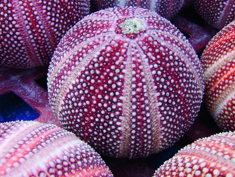
Coorssen’s team primarily uses release-ready cortical vesicles from unfertilised sea urchin eggs in their work on regulated exocytosis.
Maturing oocytes, eggs and the early embryos of sea urchins have been used as key model systems for well over a century, providing critical cellular and molecular insights into mechanisms underlying fertilisation, development, calcium signalling, the immune response, cell cycle regulation and regulated exocytosis.
“It is a highly conserved system in which the docked, release-ready and late calcium-triggered steps of exocytosis are isolated and can be quantitatively assessed,” said Coorssen. “We use different proteomic and lipidomic approaches to dissect what’s going on in the mechanisms of triggered release/regulated exocytosis.”
Despite decades of research, the calcium-triggered fusion mechanism involved in exocytosis remains poorly defined at the molecular level. Understanding this mechanism is a critical requirement to targeting this pathway for rational drug development.
Like the majority of eukaryotic cells, sea urchin eggs contain secretory vesicles, but in this case, more importantly, stage-specific, release-ready cortical vesicles. These membrane-bound organelles fuse with the plasma membrane to release their contents into the extracellular space. This highly conserved and fundamental cellular process of regulated exocytosis forms the basis of a diversity of functions including fertilisation, intracellular trafficking, wound healing and neurotransmission.
As might be expected of the close functional relationship to mammalian systems, there is also a very high degree of genetic conservation between sea urchins and humans.
“The sea urchin genome has been sequenced,” added Coorssen. “There is high genetic conservation from sea urchins to humans, which makes it a very reliable model system in that respect. Critical and fundamental mechanisms are highly conserved or they cease to exist. Such is the nature of most critical molecular mechanisms.”
A chemical proteomics approach
One aim of the work in Coorssen’s team is to develop a list of key players involved in the mechanism of regulated exocytosis and assign them to direct or modulatory roles.
They have been using different biophysical, (bio)chemical, proteomic and lipidomic approaches to identify the critical players and their roles.
“We have a lot of tools to link the physiology and the proteomics. It’s functional proteomics, and the reason we use this model system is that it enables us to link as tightly as possible the quantitative functional and the quantitative molecular assays. That’s the only true route to dissecting mechanism.”
These tools include two chemical proteomic approaches, one is based on differential protease effects.
“In the past, one of the techniques we used was differential sensitivity to proteases,” recalled Coorssen. “We were able to proteolytically remove key proteins that were suggested to be essential to the fusion mechanism and demonstrate quantitatively that we’d removed the critical cytosolic domains of those proteins.
“Yet the calcium-triggered fusion mechanism still worked. As we did this quantitatively, we could confirm that these components were clearly important, but were not acting in the way they were claimed to according to the major hypothesis in the field at the time regarding the protein machine that was believed to drive fusion. They’re working upstream of triggered fusion. Thus, the docking and priming roles we postulated instead, years ago, have since been confirmed by others, in other secretory cell types.”
The researchers are now integrating this approach using broad-spectrum proteases with another approach using selective inhibitors of different kinases and phosphatases to test for effects on triggered release. In this way they are targeting the identification of critical alterations to the phosphoproteome of secretory vesicle membranes, including parallel validation in synaptic preparations.
“There are multiple things going on at a biological membrane and the membrane is thus chock-full of proteins that are not at all involved in your mechanisms of interest. One of the things the protease treatment does is remove a lot of those yet leave the triggering and release mechanisms intact, so any proteins that are removed must not be critical to mechanism. One can, to some extent, be rid of ‘background’.
“The cortical vesicle model thus enables us to home in on critical proteins to better refine subsequent validation experiments in mammalian secretory cells that might otherwise take years of research funding.”
The other chemical proteomic approach involves thiol-labelling. Using this technique the researchers have identified two classes of reagents that act in opposite ways - one promotes the calcium sensitivity and kinetics of vesicle release and the other inhibits it.
“Using multiple fluorescent reagents we are now simultaneously modifying function while labelling the proteins involved,” explained Coorssen. “One simply can’t do such direct, coupled, quantitative analyses and identification of critical proteins with any other model system. Naturally, validation in synaptic and other clinically relevant mammalian secretory cells are also part of this long-term and well-defined research program.”
The membrane proteome
High-resolution, gel-based analyses have enabled Coorssen’s team to dissect the cortical vesicle membrane proteome. Now that they have the proteome they are studying it to understand critical underlying molecular mechanisms.
“Part of the reason we do gel-based proteomics is because it directly delivers that critical quantitative aspect, as well as the necessary deeper analysis of protein species, in parallel, across several replicate samples,” said Coorssen.
Using gel electrophoresis is also helping to identify critical protein interactions. Membrane protein complexes involved in vesicular docking and in triggering fusion have been resolved and, together with chemical proteomic approaches, this will enable the researchers to define the components that make up the physiological fusion machine of regulated exocytosis.
This highly quantitative approach, directly coupling the functional and molecular assays, enables the researchers to analyse protein and lipid components of the system and work out which components, and which interactions, are essential to both setting up and driving the fusion of vesicles to the membrane.
Lipidomic analyses
In parallel, Coorssen and colleagues have also been equally interested in the role of lipids in the exocytotic pathway. His team was the first to identify cholesterol as a critical component of the exocytotic mechanism.
“Cholesterol is a central conserved component of exocytosis and of the final triggered fusion process, but that exact function will be difficult to fully dissect outside of the cortical vesicle, particularly with regard to membrane curvature contributions,” said Coorssen, “and that is also true of other such components. However, time-resolved electrophysiological analyses of the fusion pore are pushing the boundaries in this respect - the coupled, quantitative function-molecular analytical approach.”
In an attempt to better define the role of cholesterol, Coorssen’s team has refined automated, high-performance, thin-layer chromatographic analyses as a quantitative approach to lipidomics. Using this technique they have shown that cholesterol is directly involved in the triggered membrane fusion mechanism.
They have also shown that reducing the cholesterol concentration in different vesicle membranes, using multiple strategies such as chemical extraction and sequestration, enzymatic reduction, and inhibition of cholesterol biosynthesis, adversely affects fusion. This approach has also identified other critical lipids, such as phosphatidylethanolamine and phosphatidylserine, and firmly established that still others (ie, polyphosphoinositides) actually have upstream modulatory roles during docking and priming.
A quantitative approach
Resolving the fundamental molecular mechanisms underlying the process of vesicular release - understanding and describing the critical docking, priming, triggering, and fusion steps of this essential cellular process - will enable the rational targeting of this pathway in the treatment of a broad range of disorders. Many of these are serious and growing healthcare burdens, such as the neurodegenerative, diabetes and related disorders.
“If you don’t do thorough quantitative work you’re never going to have more than a cartoon of your mechanisms,” said Coorssen. “And a cartoon isn’t good enough for identifying, for instance, critical targets for drug development. You need to know that the target you’re after is genuinely a target of interest.”
**********************************************************
Lorne conference line-up
Here’s the line-up for the Lorne conferences for 2014, to be held at Mantra Lorne on the Victorian south coast.
19th Lorne Proteomics Symposium
February 6-9
http://www.australasianproteomics.org/lorne-proteomics-symposium-2014/
39th Lorne Conference on Protein Structure and Function
February 9-13
http://www.lorneproteins.org/
26th Lorne Cancer Conference
February 13-15
http://www.lornecancer.org/
35th Lorne Genome Conference
February 16-19
http://www.lornegenome.org/
Lorne Infection and Immunity
February19-21
www.lorneinfectionimmunity.org
**********************************************************
Personality influences the expression of our genes
An international research team has used artificial intelligence to show that our personalities...
Pig hearts kept alive outside the body for 24 hours
A major hurdle for human heart transplantation is the limited storage time of the donor heart...
Breakthrough antibiotic for mycobacterial infections
The antibiotic candidate, named COE-PNH2, has been optimised to target Mycobacterium...


